Q27 of 30 Page 1

Draw a schematic sketch of pBR322 plasmid and label the following in it:

(a) Any two restriction sites.


(b) Ori and rop genes.


(c) An antibiotic resistant gene.


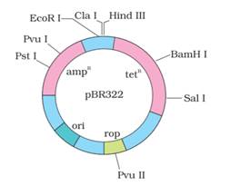
Here is a schematic sketch of pBR322 plasmid:


a) Restriction sites Hind III and EcoRI are shown.


b) The Ori and rop genes are shown.


c) Antibiotic resistant gene TetR and AmpR are shown as well.


More from this chapter

All 30 →
25

Given below is the representation of amino acid composition not the relevant translated portion of β-chain of haemoglobin, related to the shape of human red blood cells


(a) Is this representation indicating a normal human or a sufferer from certain related genetic disease? Give reason in support of your answer.


(b) What difference would be noticed in the phenotype of the normal and the sufferer related to this gene?


(c) Who are likely to suffer more from the defect related to the gene represented the males, the females or both males and females equally? And why?


26

By the end of 2002 the public transport of Delhi switched over to a new fuel. Name the fuel. Why is this fuel considered better? Explain.

28

Explain the carbon cycle with the help of a simplified model.

OR


(a) Explain how does a primary succession start on a bare rock and reach a climax community?


(b) Explain how does the algal bloom eventually choke the water body in an industrial area?


29

The following is the illustration of the sequence of ovarian events (a − i) in a human female


(i) Identify the figure that illustrates ovulation and mention the stage of oogenesis it represents.


(ii) Name the ovarian hormone and the pituitary hormone that have caused the above mentioned event.


(iii) Explain the changes that occur in the uterus simultaneously in anticipation.


(iv) Write the difference between ‘c’ and ‘h’.


(v) Draw a labeled sketch of the structure of a human ovum prior to fertilization.


OR


(a) How does the megaspore mother cell develop into 7-celled, 8 nucleate embryo sac in an angiosperm?


(b) Draw a labeled diagram of a mature embryo sac.